Wednesday, June 14, 2017
An Update on Avalanche Biotechnologies A Potential Longer Lasting Wet AMD Treatment
Wednesday, May 31, 2017
NeoVista Epi Retinal Strontium 90 Treatment for AMD Update 4
To bring you up-to-date, I began following NeoVista in February 2007, writing an initial piece describing the procedure and initial clinical results. I also asked a series of questions of management and printed their responses. That first piece was:
NeoVista Epi-Retinal Strontium 90 Treatment for Wet AMD
In July 2007, the company announced the initiation of the CABERNET clinical study, and I posted an update, including more questions about the study to management. This piece was:
NeoVista Epi-Retinal Strontium 90 Treatment for AMD Update
NeoVista Epi-Retinal Strontium 90 Treatment for AMD: Update 2
And, the final update (until now) was written following the 2008 Retina Society Meeting in September 2008. This included 18-month data from the Phase II feasibility study, and concluded with the statement: “With the continued promise of these Phase II trial results, NeoVista continues to enroll patients in the company's pivotal trial, CABERNET. CABERNET is a multicenter, randomized, controlled study that will enroll 450 subjects at 45 sites worldwide, evaluating the safety and efficacy of NeoVista's epiretinal brachytherapy delivered concomitantly with the FDA-approved anti-VEGF therapy Lucentis (ranibizumab) versus Lucentis alone.”
NeoVista Epi-Retinal Strontium 90 Treatment for AMD: Update 3
This new update (Update 4) will attempt to bring you up-to-date on both the clinical trials underway, as well as provide a few quotes from the press releases announcing the patient treatments upon commercialization of the device in Europe.
Commercialization in Italy
From the press release about the first patients treated in Pisa, Italy, announced November 12, 2009:
The first VIDION patients were treated by Dr. Stanislao Rizzo, from the S. Chiara Hospital,Azienda Ospedaliera Universitaria Pisana, Pisa, Italy, who said, “We are very excited to be the first hospital to perform this innovative one time treatment procedure, which can help to treat a devastating disease that otherwise requires chronic treatment for an indefinite period of time. NeoVista’s targeted epimacular brachytherapy treatment may provide us the ability to improve vision by offering a distinct mechanism of action that affects multiple disease pathways unlike the conventional anti-VEGF therapy, and may dramatically change the patient’s quality of life by eliminating frequent eye injections. This promising treatment is a cost-effective alternative to treat neovascular AMD,” continued Dr. Rizzo. “Monthly injections can become quite expensive for our health care system, whereas a single procedure will potentially allow our specialists to treat more patients and bring down the costs associated with ongoing treatments.”
Commercialization in the United Kingdom
From the press release about the first patient treatments in London, UK, announced November 16, 2009:
The new device is initially being introduced in 15 hospitals across the UK as part of a large clinical trial called MERLOT, where it will be used in patients whose current standard of care treatment involves regular injections of a drug into the eye to control their condition (up to one injection every month, indefinitely). Although the treatment is initially only available at select hospitals, it is anticipated that the number of sites using the device will increase quickly, to provide nationwide availability.
The MERLOT trial has been recently awarded portfolio status by The National Institute for Health Research (NIHR) Comprehensive Clinical Research Network (CCRN). The CCRN was created as part of the government’s research and development strategy, “Best Research for Best Health” to provide a world-class infrastructure for clinical trials in all areas of disease and clinical need within the NHS in UK.
Mr Tim Jackson, a Consultant Eye Surgeon at King’s College Hospital, who is the lead investigator for MERLOT said: “This is a relatively straightforward operation and the published results are very impressive. My experience in our own trials of this device has also been encouraging and it is a big step forward to be able to offer patients this new treatment throughout the UK, within a large randomized controlled clinical trial.”
(For more on MERLOT, see the clinical trial section below.)
Commercialization in Germany
NeoVista, Inc. announced today the first commercial utilization of Epimacular Brachytherapy in Germany. Epimacular Brachytherapy is performed using the VIDION ANV Therapy system and is being offered as an adjunct therapy to anti-VEGF injections for the treatment of neovascular age-related macular degeneration.
John N. Hendrick, President and CEO of NeoVista commented, "Today is an extraordinary day for NeoVista, our local business partner, OctreoPharm Vertriebs GmbH, and the multitude of patients who suffer from this debilitating and life-altering disease. The burden of wet AMD to those afflicted with the disease, and their caregivers, is enormous."
Professor Gisbert Richard, Professor and Head of the Ophthalmology Department in the University Medical Center of Hamburg-Eppendorf, stated, "Our highly qualified medical staff is quite pleased to now be able to offer another therapeutic option to patients suffering from wet AMD - especially those patients who require frequent anti-VEGF injections." Multiple studies have demonstrated that injections alone are not able to effectively treat this disease in a large portion of the population."
Mr. Hendrick added, "The German health care system presents a welcome opportunity for NeoVista to demonstrate the effectiveness of this emerging technology. I believe the use of Epimacular Brachytherapy in the treatment of wet AMD will continue to gain momentum in Germany, and in many other countries, as the rising cost of health care, on a global basis, encourages more and more patients to continue seeking out additional or alternate therapies."
The NeoVista approach to treating wet AMD delivers a focused dose of strontium 90 beta radiation directly to the back of the eye, without damaging the adjacent healthy retinal vasculature. Importantly for patients, the systemic exposure to radiation is minimal and highly controlled to a local area. The effective dose to the entire body from NeoVista's device is less than that from a typical chest x-ray.
Clinical Trial Studies
Pilot Studies:
NVI-068
The NVI-068 trial was a study of subjects treated with a single dose of 15 or 24 Gy epimacular brachytherapy. Safety parameters evaluated included incidence and severity of ocular adverse events identified by slit lamp and indirect ophthalmoscopic examination, fluorescein angiography, and optical coherence tomography. Patients will be followed in this trial for 3 years to evaluate safety. (24 month data has been presented.)
NVI-111
The NVI-111 trial was a study of subjects treated with a single dose of 24 Gy epimacular brachytherapy and two injections of bevacizumab (1.25 mg). Subjects received one injection prior to surgery (10±4 days) or at the time of surgery and the second injection at Month 1. Subjects were re-treated with bevacizumab per the investigator’s discretion at follow-up visits. Safety parameters evaluated included incidence and severity of ocular adverse events identified by slit lamp and indirect ophthalmoscopic examination, fluorescein angiography, and optical coherence tomography. Patients are being followed in this trial for 3 years to evaluate safety. (36 month data has been presented and submitted for Peer Publication)
Feasibility Study:
MERITAGE I
MERITAGE I is a multi center international feasibility study designed to decrease the burden of treatment, it is fully enrolled (n=53) and is ongoing. The study is designed to evaluate the safety and efficacy of epimacular beta radiation therapy in patients that require persistent frequent anti-VEGF therapy to treat Wet AMD. It is planned for two sites, one in the US and one in the UK. (12 month data has been presented and a manuscript is being prepared for Peer Publication)
Pivotal Studies:
CABERNET
With the continued promise from the above two Phase II trial results (NV-068 and NV-111), NeoVista has finished enrollment in the company’s pivotal trial, CABERNET. CABERNET is a multicenter, randomized, controlled study that has enrolled 492 subjects at 45 sites worldwide, evaluating the safety and efficacy of NeoVista’s epiretinal brachytherapy delivered concomitantly with the FDA-approved anti-VEGF therapy Lucentis® (ranibizumab) versus Lucentis alone. (Initial results will be reported in Q4, 2011)
Meritage II
A pivotal study for FDA approval treating chronic wet AMD patients. (This study idea has been abandoned)
Special Population/Reimbursement Studies:
Pinot
A feasibility study treating a specific vascular tumor. Begun in Q3 2009, and still underway..
Merlot
An investigator-sponsored study in the UK; It is a head-to-head comparison of NeoVista epimacular beta radiation therapy and standard-of-care treatment with Lucentis®, begun in Q4 2009 and >40% enrolled.
Thursday, May 18, 2017
Avastin Update 5 NIH Considers Trial Comparing Lucentis and Avastin
NIH Considers Trial Comparing Lucentis and Avastin
By: Dru Thomas, Managing Editor, Ophthalmic Market Perspectives
Amidst a controversy about treatment prices, the NIH is considering funding a study to compare results of AMD therapies with Lucentis and with Avastin.
Lucentis, Genentech's long-awaited anti-angiogenic therapy for age-related macular degeneration (AMD), received FDA regulatory approval on June 30, and the Company immediately shipped $10 million worth of the drug. However, the rosy prospects for Genentech and for wet AMD sufferers took a more convoluted path in mid-July. The US National Institutes of Health (NIH) indicated it was considering funding a clinical study that compares the efficacy of Lucentis with that of Avastin, Genentech's anti-cancer agent that has also shown very positive results in off-label use against wet AMD.
In terms of both science and medical advancement, the two drugs might seem to present a win-win situation, but the reality of the marketplace creates a decidedly more complicated scenario. Genentech, a biotech powerhouse, has created two drugs that may each offer a quantum leap forward in successful AMD treatment, yet the Company faces a unique situation in which its own highly-successful anti-cancer drug may undercut the revenue stream of its proven anti-AMD agent. The crux of the issue is money.
Genentech developed Avastin and received FDA regulatory approval for its use as a systemic treatment for colorectal cancer. Avastin is central to the Company's revenues-both present and future-and the drug is currently involved in scores of trials concerning as many as 25 types of tumors. It is also used off label for some cancers.
Recognizing that the anti-angiogenic Avastin molecule might have potent applications in AMD, Genentech created Lucentis by deriving a fragment from the larger Avastin molecule. This smaller fragment was genetically engineered to create a higher binding affinity than Avastin and to penetrate the retina more effectively when administered by intravitreal injection.
As Lucentis made its way through FDA clinical trials, some doctors were impatient to offer real help to their AMD patients. Buoyed by promising reports regarding the closely-related drugs, they began treating wet AMD patients with intravitreal injections of small amounts of Avastin, an off-label use of the drug. As evidence of positive outcomes accumulated, the practice spread.
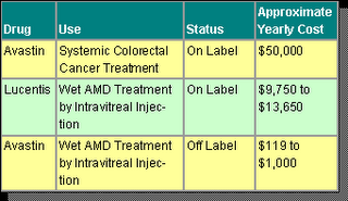
Now, with Lucentis available in the marketplace, pricing for AMD therapy is causing quite a stir. Clearly, Genentech has invested significant R&D dollars in both Avastin and Lucentis, including years of testing and clinical trials. Avastin is currently sold as an intravenous treatment and commonly costs a colorectal cancer patient approximately $50,000 per year. Lucentis is injected intravitreally and typically costs an AMD patient between $9,750 and $13,650 per year. But here is the rub-when Avastin is used in the small dosages appropriate for intravitreal injection in AMD patients, the yearly cost is less than $1,000.

There is an enormous difference in price between these two AMD treatments. As health care providers, insurers and public health systems in many nations feel the pressures of mounting health care costs, there is clearly incentive for further examination of AMD therapy using Avastin.
Meanwhile, the NIH has given no timetable for its decision about funding a comparative study, but if the study takes place and if Avastin is proven to be as effective as Lucentis, findings may jeopardize Lucentis' future earnings. However, these are big "ifs," and any study will take years to conduct. At present, doctors may choose between two Genentech drugs that appear to be remarkably effective against wet AMD. One, Lucentis, is used on label and is eligible for reimbursement; the other, Avastin, involves non-reimbursable, off-label use that might increase a doctor's liability if anything goes wrong. For the time being, Lucentis has a clear edge in the marketplace.
(Reprinted with permission from Market Scope. For information on subscribing to Ophthalmic Market Perspectives, please go to www.marketscope.com.)
Author’s Note on Avastin
Since the original posting on January 31, 2006, I have now added eight updates on this important drug for treating age-related macular degeneration. In addition to the posting you are reading, here is a listing (with links) to the others:
Avastin: A New Hope for Treating AMD (January 2006)
Avastin Update: Medicare not Likely to Cover its Use (March 2006)
Avastin Update II: AAO supports Medicare Coverage for Off-label Avistan Use (April 2006)
ARVO 2006: A Further Update on Both Avastin and Lucentis for Treating AMD (May 2006)
Avastin/Lucentis Update 4: FDA Approves Lucentis for Treating Wet AMD (July 2006)
Avastin/Lucentis Update 6: Latest Results Published in NEJM and Another Call for a Trial Between Them (October 2006)
Avastin/Lucentis Update 7: BREAKING NEWS – NEI/NIH Will Fund Comparative Study (October 2006)
Friday, April 28, 2017
Stem Cells in Ophthalmology An Update AstraZeneca Joins the Fray
Wednesday, March 8, 2017
Iluvien Update 2 New Safety and Efficacy Data Presented at ARVO
Earlier this week, Alimera Sciences and pSivida Corporation jointly announced that one of the FAME Study investigators had presented on a subset of the FAME Study data at the ARVO Meeting in Fort Lauderdale, and that the company (Alimera) plans to submit this new subgroup data to the FDA in support of its New Drug Application. Iluvien is licensed by pSivida to Alimera Sciences, Inc.
Here are the details:
Alimera's New 36-Month Safety and Efficacy Results From the Phase 3 Fame Study of Iluvien in Patients With Diabetic Macular Edema Presented at the 2011 Arvo Annual Meeting
Alimera Plans to Submit This New Data to the FDA in Support of Its Pending New Drug Application
Alimera Sciences, Inc. announced that positive new data from the completed FAME Study of Iluvien were presented at the 2011 Association for Research in Vision and Ophthalmology (ARVO) Annual Meeting. The new data showed that 33.6% of patients in Trial A (p<0.001) and 42.4% of patients in Trial B (p<0.001) were observed achieving best corrected visual acuity (BCVA) improvement of 15 letters or more from baseline at month 30 in the identifiable subgroup of patients diagnosed with diabetic macular edema (DME) for three years or more at baseline. The new data were presented by Dr. Andrew N. Antoszyk, one of the FAME investigators and a practicing retina specialist at Charlotte Eye, Ear, Nose and Throat Associates in Charlotte, N.C.
The new data, presented by Dr. Andrew N. Antoszyk, analyzed the subgroup of patients who had been diagnosed with DME for three or more years at entry of the FAME Study (which comprises over 50% of patients in the Study).
The FAME Study consisted of two three-year, Phase 3 pivotal clinical trials (Trial A and Trial B) to assess the safety and efficacy of Iluvien in the treatment of DME. The 956 patients in the trials were randomized to receive either high dose Iluvien, low dose Iluvien or control treatment. The primary endpoint for efficacy in the trials was the difference in the percentage of patients whose BCVA improved by 15 or more letters from baseline on the Early Treatment Diabetic Retinopathy Study (ETDRS) eye chart at month 24 between the treatment and control groups.
As previously reported, the pre-specified primary endpoint for the FAME Study was met for the low dose Iluvien in both Trial A and Trial B. Based on these data, Alimera submitted a New Drug Application (NDA) to the U.S. Food and Drug Administration (FDA) on June 29, 2010 for approval of the low dose Iluvien. Therefore, only the low dose data is presented and discussed here.
In February 2011, Alimera presented positive results from the full patient population at month 36 of the FAME Study in Trial A (28.4%) and Trial B (29.0%) with demonstrated improvement in BCVA of 15 letters from baseline. Statistical significance was seen in both trials as late as month 33 with Trial A at 28.4% (p=0.042) and Trial B at 29.6% (p=0.046).
Dr. Antoszyk's ARVO presentation on May 3rd included additional data from a subgroup of study patients that was identifiable prior to administration of Iluvien. This subgroup reflected the duration of DME at baseline and across all patients randomized, with a median duration of DME at baseline of three years.
In the data reported for this subgroup at 36 months in Trial A, 31.8% of patients treated with Iluvien experienced an improvement in best corrected visual acuity (BCVA) of 15 or more letters from baseline compared with 13.6% of those in the control group (p=0.010), for a net benefit of Iluvien versus control of 18.2%. In Trial B, 36.4% of Iluvien patients in this subgroup experienced improvement of 15 or more letters compared to 13.2% of control patients (p= 0.004), for a net benefit of Iluvien versus control of 23.2%. On a combined basis for both Trials A and B, at three years the net benefit of Iluvien compared to control reported for patients in the subgroup was 20.6%, more than double that seen for the full patient population (9.8%).
In the subgroup, peak efficacy was seen at month 30, with 33.6% of Iluvien treated patients in Trial A gaining 15 or more letters in BCVA compared to 10.2 % of control (p < 0.001) and 42.4% of Iluvien treated patients in Trial B gaining 15 or more letters in BCVA Trial B compared to 11.3% of control (p< 0.001).
Consistent with the full patient population in the FAME Study, approximately 75% of the patients in this subgroup treated with Iluvien were reported to have received only one Iluvien insert over the 36 month study.
There was no statistically significant difference in BCVA improvement in the subgroup of patients with less than three years' duration of DME at entry compared to control.
"Throughout the FAME Study, Iluvien has shown significant potential for patients suffering with DME. This new data is particularly exciting with 34% of patients who've had DME for three years or more gaining three lines of vision after therapy," said Dr. Antoszyk. "If regulatory approval of Iluvien is obtained, we will be able to offer an additional option in the form of a long-term treatment to our patients who are dealing with this devastating disease."
Data for the subgroup was gathered from 536 patients who had been diagnosed with DME for three years or more and 416 patients who had been diagnosed with DME for less than three years. Alimera will provide these additional data in its response to the Complete Response Letter issued by the FDA in December 2010.
Safety was assessed among those patients within the subgroup who were treated with Iluvien in the study. Intraocular pressure (IOP) increases to 30 millimeters of mercury (mmHg) or greater at any time point were seen in 14.8% of these patients by month 36, compared to 18.3% in the full Iluvien treated patient population. By month 36, 5.3% of these patients had undergone an incisional surgical procedure to reduce elevated IOP, compared to 4.8% in the full patient population. The incidence of cataracts among patients with a natural lens in their eye at the start of the study was 86% at month 36, with 85% undergoing a cataract operation, compared to 80% and 74.9%, respectively, in the full patient population.
"We are pleased that this identifiable subgroup shows even greater benefit to risk than the full patient population through month 36 of the study, thereby further improving Iluvien's profile," said Dan Myers, Alimera's president and CEO. "This data spotlights the benefit that Iluvien, if approved, could bring to the patient population that retinal specialists are targeting for its use. We believe this data will be very valuable to the treatment of DME going forward."
Paul Ashton, president and chief executive officer of pSivida, said, "We are very pleased with the efficacy and safety results through month 36 in patients with chronic DME. This subgroup comprised a majority of patients in the FAME Study. We look forward to Alimera's filing of this data with the FDA in connection with the NDA for Iluvien."
About the FAME Study
Alimera conducted two 36-month, Phase 3 pivotal clinical trials (collectively known as the FAME Study) for Iluvien involving 956 patients in sites across the United States, Canada, Europe and India to assess the efficacy and safety of Iluvien with two doses of the corticosteroid fluocinolone acetonide (FAc), a high and low dose, for the treatment of DME. The primary efficacy endpoint for the FAME Study was the difference in the percentage of patients whose best corrected visual acuity improved by 15 or more letters from baseline on the ETDRS eye chart at month 24 between the treatment and control groups. The study concluded in September 2010 with the final patient visit at the three-year data point.
Following its NDA submission to the FDA, Alimera submitted a Marketing Authorization Application to the Medicines and Healthcare products Regulatory Agency in the United Kingdom. Applications have also been submitted to regulatory agencies in Austria, France, Germany, Italy, Portugal and Spain. Based upon the analysis of the FAME Study, all filings included the 24-month data. The FDA, in a December 2010 Complete Response Letter, requested further information including the month 36 data from the FAME Study.
About DME
DME, the primary cause of vision loss associated with diabetic retinopathy, is a disease affecting the macula, the part of the retina responsible for central vision. When the blood vessel leakage of diabetic retinopathy causes swelling in the macula, the condition is called DME. The onset of DME is painless and may go undetected by the patient until it manifests with the blurring of central vision or acute vision loss. The severity of this blurring may range from mild to profound loss of vision. The Wisconsin Epidemiologic Study of Diabetic Retinopathy found that over a 10-year period approximately 19% of people with diabetes studied were diagnosed with DME. As the population of people with diabetes increases, Alimera expects the annual incidence of diagnosed DME to increase, as well.
Sunday, February 12, 2017
Stem Cells in Ophthalmology Update 24 Current Tables Now Online
Updated December 31, 2015.
Sunday, January 8, 2017
CATT Study Update 4 Avastin vs Lucentis Study Ready to Roll
In particular: What Is Neovascular AMD and Why Is the CATT Study Important?
This section provides an explanation of what AMD is and how it is treated, along with some information about the CATT Study, its design and aims.
The Study’s primary aim is to evaluate the relative safety and efficacy of treatment of subfoveal AMD with both Avastin and Lucentis, determine an appropriate dosing schedule and, to see if there is any clinical difference between the two drugs. Some of the secondary aims include:
- Determination of the number of treatments required at 1 and 2 year periods
- Determination if either or both drugs provide a 3-line change in visual acuity (15 letters on ETDRS chart)
- Changes in subretinal and intraretinal fluid on OCT examination
- Changes in lesion size on fluorescein angiograph examination
- Incidence of any complications of treatment (endophthalmitis, retinal detachment, cataract, uveitis)
- Incidences of other adverse effects
- Comparison of the cost of treatment over two years
There are two links within the document: one describes the eligibility requirements, while the second is a 258 page Manual of Procedures, which contains everything your ever wanted to know about the CATT Study.
The only changes I noted from the original information I have provided are that enrollment is now scheduled to begin January 1st, and the number of clinical sites has been reduced to 44. The location of the 44 sites is not provided on the website (yet), but hopefully will be included soon.
In any event, I have a prior list of 45 sites which presumably contains all of the sites scheduled to participate. Anyone wishing to know what sites are participating in their states can email me and I will be happy to provide a list for that state. (My email link is shown in the sidebar.)
Wednesday, December 21, 2016
Stem Cells in Ophthalmology Update 15 Wills Eye Joins ACT’s Clinical Trials for Dry AMD Using Embryonic Stem Cell derived RPE
Breaking News – as of January 19th, Wills Eye had been added to ACT’s clinical protocol for treating Dry AMD with embryonic stem cell-derived RPE cells, and was actively recruiting patients.
Saturday, October 8, 2016
Gene Therapy in Ophthalmology Update 12 First Gene Therapy Approval on the Horizon
Thursday, September 29, 2016
AMD Update 6 An Overview of New Treatments for Dry AMD
I have previously written about the potential for the use of lasers to treat dry AMD by Iridex, but that attempt did not prove successful. I have also written about the Ellex 2RT (retinal regeneration) program that might hold potential in the early intervention in dry AMD. Links to both of these writeups are shown at the end of this posting.
With about 80% to 90% of newly diagnosed AMD cases being of the dry variety, and with no effective treatment currently in use, it is important to track the developmental work underway in this area. This review appeared in the November 2009 issue of Retinal Physician, and with the permission of the magazines publishers, here is the complete writeup:
Preclinical and Phase 1 Drugs in Development for Dry AMD: An Overview
Retinal Physician, November 2009
Philip J. Rosenfeld, MD, PhD ● John Legaretta, BFA
Philip J. Rosenfeld, MD, PhD, is professor of ophthalmology at the Bascom Palmer Eye Institute at the University of Miami Miller School of Medicine in Florida. He receives significant research support from Potentia and Alexion, and he has a minimal advisory relationship with Potentia. John Legarreta, BFA, is a medical student at the School of Medicine and Biomedical Sciences, State University of New York at Buffalo. He has no financial interest in any products mentioned in this article. Dr. Rosenfeld can be reached at prosenfeld@med.miami.edu.
The vast majority of AMD patients have the nonexudative or dry form of the disease, characterized by a constellation of clinical features, including drusen, disturbances of the retinal pigment epithelium (pigment clumping and/or dropout), and geographic atrophy (GA) of the macula. As defined by the Age-Related Eye Disease Study (AREDS), the severity of AMD can be classified into three categories: early, intermediate, and advanced.
[Editors Note: For a breakdown on the number of people in the three categories noted above in both dry and wet AMD in 2007, as estimated by Market Scope, please see the link to the table shown at the end of this posting.]
While drusen alone, particularly those of smaller size, do not seem to be associated with vision loss, at least one large druse measuring 125 ฮผm in diameter is sufficient for the diagnosis of intermediate AMD. Dry AMD may remain static or progress slowly to produce a greater number and distribution of drusen with areas of GA. The increase in size or area of drusen or pigment abnormalities (focal hyper- or hypopigmentation of the retinal pigment epithelium [RPE]) predicts the likelihood of developing vision-threatening lesions in AMD, which include central GA and neovascularization, the advanced forms of AMD.(1)
CURRENT TREATMENT OPTIONS FOR DRY AMD
Antiangiogenic therapies have been developed to treat wet AMD. While drugs such as ranibizumab (Lucentis, Genentech) and bevacizumab (Avastin, Genentech) have revolutionized the care of patients with neovascular AMD, under the best of circumstances, treatment converts the neovascular form of AMD back to dry AMD. There is no evidence to suggest that these antiangiogenic drugs have any beneficial effect on the underlying degenerative process known as dry AMD. Currently, there is no proven drug treatment for dry AMD; however, the cessation of smoking and treatments based on nutritional recommendations and supplements can slow disease progression. Nutrient-based treatments for AMD were evaluated in the AREDS trial.(2)
TARGETING THE CAUSE OF AMD
The overall goal of treatment for dry AMD is to target the underlying cause of the disease and halt, or at least slow, the loss of vision. This approach has been hampered by two major issues. First, there are no reliable in vitro systems for testing the efficacy of any drug for dry AMD, and second, no true animal model exists for AMD. A well-developed macula is only found in primates and birds, and while numerous attempts have been made to develop nonprimate models for AMD, and these models highlight various pathological features of human AMD, none of these animal models truly replicates the disease process seen in humans. The only model that may be useful for potential drug testing is the naturally occurring monkey colonies that have been found to develop drusen.(3)
The second issue that has hampered drug development is the uncertainty surrounding the best molecular pathway to target for the treatment of dry AMD. However, several different strategies have evolved. These strategies have targeted three major therapeutic areas of investigation: preservation of photoreceptors and the RPE (neuroprotection), prevention of oxidative damage, and suppression of inflammation. Each strategy is supported by varying degrees of scientific evidence and will have to await validation based on clinical trial outcomes.
CLINICAL TRIAL ENDPOINTS IN DRY AMD
The most obvious study endpoint for dry AMD therapies would be the preservation of visual acuity; however, studies using visual acuity as an endpoint will take many years to complete. To decrease the time required to show a benefit from a drug, surrogate endpoints have been developed that might indicate a positive outcome without waiting the years required to show visual acuity benefit.
One surrogate endpoint is the prevention of disease progression from dry to wet AMD. This endpoint was first used in the study investigating anecortave acetate (Retaane, Alcon) for the treatment of dry AMD. While the drug failed to prevent progression of dry to wet AMD, the study demonstrated the feasibility of this study design. Another strategy is to assume that a treatment for dry AMD might also affect the underlying stimulus for neovascularization in wet AMD. If true, then a potential endpoint might be to demonstrate that a drug for dry AMD is able to decrease the need for retreatment with antiangiogenic therapy in wet AMD or improve the visual acuity outcome. This study design has not been tested.
A feature of dry AMD that could serve as a surrogate endpoint is the area of drusen in the macula. While drusen area as measured by fundus photography has already been explored as an endpoint in the failed laser-to-drusen trials, (4-6) the change in drusen volume in response to pharmacotherapy is a novel clinical trial endpoint that has not been explored previously. Spectral-domain optical coherence tomography has the potential to reliably and reproducibly identify drusen in the macula and provide truly automated volume quantification. The most likely surrogate clinical trial endpoint, based on a symposium held in Washington, DC, and sponsored by the National Eye Institute and the Food and Drug Administration, is an endpoint that assesses a drug’s effects on the growth of GA, since GA is a feature of dry AMD that directly causes loss of photoreceptors and the RPE. (7)
DRUGS TO PROMOTE SURVIVAL OF PHOTORECEPTORS AND THE RPE
No matter what the underlying cause of AMD, drugs that can preserve viable photoreceptors and maintain the RPE should preserve vision. One strategy to promote survival of photoreceptors and the RPE is to protect cells against ischemia and improve the choroidal circulation in patients with dry AMD. Two studies are currently using this strategy. In Europe, an ongoing multicenter, randomized, placebo-controlled study is investigating the use of an offlabel, generic drug known as trimetazidine (Vastarel MR, 35 mg tablet), a drug currently used for the treatment of angina pectoris. Trimetazidine improves myocardial glucose utilization by stopping fatty acid metabolism, and it is considered to have cytoprotective effects in ischemic conditions. Other uses for this drug include the treatment of vertigo, tinnitus, and vision loss and visual field loss due to vascular causes. The primary goal of this study is to slow the conversion of dry AMD to wet AMD.
Another drug being investigated for its vasodilatory effect is Alprostadil, also known as prostaglandin E1 (PGE1). The presumed rationale is based on the belief that improved circulation would slow the progression of AMD. This multicenter, randomized, placebo-controlled study is ongoing in Europe.
Another strategy to preserve the macular function is to prevent apoptosis by using neuroprotective agents. Ciliary neurotrophic factor (CNTF), a potent neuroprotective agent, has been shown to inhibit photoreceptor apoptosis in an animal model of retinal degeneration (8) and is being investigated as a treatment for dry AMD. Using encapsulated cell technology that permits CNTF-producing transfected cells to be implanted into the vitreous cavity, Neurotech Pharmaceuticals (Lincoln, RI) has developed a sustained-release platform that produces CNTF for a year or longer. The phase 2 study is completed and data analysis is currently under way. Other neuroprotective agents currently under investigation for dry AMD include a brimonidine tartrate intravitreal implant (Allergan, Irvine, CA) and topical tandospirone (Alcon, Fort Worth, TX).
Yet another strategy is to interfere with the normal visual cycle and preserve vision by decreasing the accumulation of toxic metabolites, such as lipofuscin and the retinal fluorophore A2E. This strategy is being pursued by Sirion Therapeutics (Tampa, FL) with the use of fenretinide - N-(4-hydroxyphenyl) retinamide - for the treatment of dry AMD. Fenretinide binds retinol-binding protein in the circulation and prevents uptake of retinol by the RPE, thus downregulating photoreceptor metabolism. The phase 2 study investigating fenretinide for the treatment of GA is fully enrolled and in its second year of follow-up.
Downregulation of photoreceptor activity is also being investigated using the drug ACU-4429 (Acucela, Bothell, WA). ACU-4429 is a small nonretinoid molecule that functions as a modulator of the isomerase (RPE65) required for the conversion of all transretinol to 11-cis-retinal in the RPE. By modulating isomerization, ACU-4429 slows the visual cycle in rod photoreceptors and decreases the accumulation of A2E. The ongoing phase 1 study has shown so far that the drug is safe and well tolerated in healthy volunteers. A phase 2 study for treatment of dry AMD is currently being planned.
A novel strategy for the preservation of photoreceptors and the RPE borrows a therapeutic strategy used for the treatment of Alzheimer's disease. An antibody against amyloid ร has completed a phase 1 study as an intravenous treatment for GA in AMD patients. This antibody, known as RN6G (Pfizer, New York, NY), was shown to decrease the amount of amyloid ร in the eye from a mouse model of AMD when given as a systemic therapy.(9) A phase 2 study is currently under way.
The therapies under investigation that seek to preserve photoreceptors and the RPE are summarized in Table 1.

(Note: I have reproduced the tables included with the article. By clicking on the tables they will open in a new window or tab in a larger more readable size. Or, go to the original online writeup for a clearer view of the table contents.)
DRUGS TO PREVENT INJURY FROM OXIDATIVE STRESS AND MICRONUTRIENT DEPLETION
In AMD, oxidative stress and the depletion of essential micronutrients are considered to be driving forces in disease progression. This disease paradigm assumes that AMD is caused by a lifelong exposure to free radicals - a byproduct of high oxygen consumption in the neural retina and RPE - combined with exposure to environmental toxins, such as those derived from smoking, in conjunction with inadequate levels of naturally occurring antioxidants. These exposures and deficits result in the accumulation of cellular debris - particularly oxidized lipids, which promote inflammation and may be directly toxic to the macular tissues - resulting in the clinical manifestations known as AMD. This paradigm is supported by epidemiologic studies showing that diets rich in antioxidants decrease the risk of AMD, while smoking was associated with an increased risk of AMD.(10)
Support for this nutrient-based paradigm was provided by the AREDS trial. This multicenter, NEI-sponsored study evaluated the effect of pharmacological doses of zinc and/or a formulation containing nutrients with antioxidant properties (vitamin C, vitamin E, and beta-carotene) on the rate of progression to advanced AMD and on visual acuity. The use of these vitamins and micronutrients reduced the risk of developing advanced AMD by about 25%.(2) The overall risk of moderate vision loss was reduced by 19% at five years. The theory of oxidative damage as a cause for AMD has also been supported by the findings that individuals have an increased risk of developing AMD if they carry a specific genetic polymorphism in mitochondrial DNA (A4917G), an organelle important for oxidative metabolism, and in nuclear DNA within the 5'-upstream region of a genetic locus important for DNA repair (ERCC6). DNA damage can be caused by oxidative stress.(11,12)
The AREDS2 trial, now under way, is designed to evaluate the effect of dietary xanthophylls (lutein/zeaxanthin) and/or omega-3 long-chain polyunsaturated fatty acids (LCPUFA), known as docosahexaenoic acid (DHA) and eicosapentaenoic acid (EPA), on the progression to advanced AMD (www.areds2.org). These micronutrients are believed to function not only as antioxidants, but also as anti-inflammatory and antiangiogenic agents, according to epidemiologic and laboratory studies. In addition, AREDS2 will investigate the effects of eliminating betacarotene and the effects of reducing zinc in the original AREDS on the development and progression of AMD.
A topical antioxidant called OT-551 (Othera Pharmaceuticals, Exton, PA) was being explored as a treatment for dry AMD. OT-551 (4-cyclopropanoyloxy- 1-hydroxy-2,2,6,6-tetramethylpiperidine HCl) is a small lipophilic molecule that readily penetrates the cornea. OT-551 is converted by ocular esterases to TEMPOL-H (TP-H), the active metabolite that is a potent free-radical scavenger that does not penetrate the cornea. In animal studies, topical therapy has resulted in excellent ocular bioavailability, with significant levels of TP-H achieved in the retina.
The drug OT-551 was shown to possess antiinflammatory and antiangiogenic properties, as well as antioxidant properties. OT-551 was also shown to protect against oxidative damage in vitro, protect against light damage in vivo,(13) suppress photoreceptor cell death in animal models, and block angiogenesis stimulated by growth factors. Based on these preclinical data, OT-551 was being investigated as a therapy for GA in AMD. This two-year, phase 2 trial, known as the OMEGA (OT-551 Multicenter Evaluation of Geographic Atrophy) study, was stopped after 18 months, due to an apparent lack of efficacy in preventing the enlargement rate of GA in AMD.
The therapies under investigation that seek to prevent injury from oxidative stress and micronutrient depletion are summarized in Table 2.

DRUGS TO SUPPRESS INFLAMMATION
Genetic association studies using different populations have shown that inflammation appears to be the driving force behind AMD.(14) In 2005, four groups identified a genetic polymorphism in complement factor H (CFH), which was associated with an increased risk of developing AMD.(15-18) The documented risk-conferring single-nucleotide polymorphism (SNP) was a thymine (T) to cytosine (C) substitution at nucleotide 1277 in exon 9, which results in a tyrosine-to-histidine change at amino acid position 402 (Y402H) of the CFH protein.
Since complement is a system of serum proteins that comprise an important arm of the innate immune system, association studies have definitively linked AMD to the immune system. Also, two independent studies reported the association of the complement factor 3 gene with AMD, (19,20) as well as the complement factor B/component 2 gene.(21) An association between the complement factor 1 gene and AMD has been reported too.(22) Less robust associations have been reported between AMD and SERPING1, which regulates the first component of complement (C1), (23) and between AMD and C7 and mannose binding lectin 2 (MBL2) loci.(24) Protective alleles associated with the complement pathway have also been reported. Two of the five CFH-related genes (CFHR1-5), which lie within the regulators of complement activation (RCA) locus on chromosome 1q32, known as CFHR1 and CFHR3, are considered to be protective against AMD.(25)
These genetic association studies would imply that inhibition of complement activation would be a reasonable strategy for the treatment of AMD. However, after a lifetime of complement-mediated damage, such a strategy might have no effect on disease progression later in life. One drug being investigated is POT-4 (Potentia Pharmaceuticals, Louisville, KY), a cyclic peptide comprised of 13 amino acids, that is derived from compstatin. POT-4 binds reversibly to complement component 3 (C3) and prevents its proteolytic activation to C3a and C3b and the subsequent release of all downstream anaphylatoxins, as well as the formation of terminal membrane attack complex. As a C3 inhibitor, POT-4 inhibits all three major pathways of complement activation. POT-4 has unique slow-release properties due to the formation of an intravitreal gel at higher doses, which should permit less frequent intravitreal injections to achieve prolonged complement inhibition.
The phase 1 dose-escalation study, known as Assessment of Safety of Intravitreal POT-4 Therapy for Patients with Neovascular AMD (ASaP), was performed on patients with advanced neovascular lesions with the intention to pursue POT-4 as a treatment for dry AMD. To date, POT-4 appears safe up to a dose of 1.05 mg, with evidence of efficacy at the higher doses.
Another complement inhibitor under investigation is eculizumab (Soliris, Alexion Pharmaceuticals), a humanized monoclonal antibody derived from a murine antihuman C5 antibody. Eculizumab specifically binds the terminal complement protein C5, thereby inhibiting its cleavage to C5a and C5b during complement activation. The strategic blockade of the complement cascade at C5 prevents the release of the downstream anaphylatoxin C5a and prevents the formation of the cytolytic membrane attack complex.
Eculizumab is FDA-approved for the intravenous treatment of another complement-mediated disease known as paroxysmal nocturnal hemoglobinuria. At the Bascom Palmer Eye Institute, we are performing a phase 2 investigation with eculizumab for the treatment of patients with dry AMD, known as the COMPLement Inhibition with Eculizumab for the Treatment of Non-Exudative Age-Related Macular Degeneration (COMPLETE) Study. Patients with GA or high-risk drusen are being randomized 2:1 to receive intravenous infusions of eculizumab or placebo.
Ophthotech's ARC-1905 (Princeton, NJ), an anti-C5 aptamer, is another complement inhibitor being tested in AMD. ARC-1905 is being administered by intravitreal injection. The phase 1 dose-escalation study was performed in combination with ranibizumab therapy for the treatment of wet AMD. Genentech/Roche are developing an anti-Complement Factor D antibody Fab (FCFD4514S), which is in a phase 2 trial. Another phase 1 study using ARC1905 for dry AMD is currently under way.
Another complement inhibitor in preclinical studies is JPE1375 (Jerini Ophthalmic, New York, NY), a small, peptidomimetic molecular antagonist against the C5a receptor, which prevents binding of C5a, thus inhibiting the biological activity of C5a. Additional complement inhibitors are being pursued in preclinical studies by several companies, but details are not yet available.
More generalized immune suppression for the treatment of dry AMD is being pursued with the use of subcutaneous glatiramer acetate (Copaxone, Teva Pharmaceuticals, Kfar- Saba, Israel), intravitreal sustained-release fluocinolone acetonide (Iluvien implant, Alimera Sciences, Alpharetta, GA), and subcutaneous sirolimus (rapamycin), a macrolide fungicide with immunosuppressive properties.
The therapies under investigation that seek to suppress inflammation are summarized in Table 3.

SUMMARY
Several different strategies are being investigated, but it will take years before we know if any of them are successful. While we await positive outcomes, these clinical trials will produce a wealth of natural history data on the progression of dry AMD and provide us with extensive experience using several different imaging modalities to track disease progression. With this experience, our clinical trial designs will improve over time and the appropriate clinical trial endpoints should become obvious. With all the drugs in development, there is a good chance that a treatment breakthrough should occur within the next decade. RP
REFERENCES
1. Ferris FL, Davis MD, Clemons TE, et al. A simplified severity scale for agerelated macular degeneration: AREDS Report No. 18. Arch Ophthalmol. 2005;123:1570-1574.
2. A randomized, placebo-controlled, clinical trial of high-dose supplementation with vitamins C and E, beta carotene, and zinc for age-related macular degeneration and vision loss: AREDS report no. 8. Arch Ophthalmol. 2001;119:1417-1436.
3. Kaidzu S, Tanito M, Ohira A, et al. Immunohistochemical analysis of aldehydemodified proteins in drusen in cynomolgus monkeys (Macaca fascicularis). Exp Eye Res. 2008;86:856-859.
4. Laser treatment in patients with bilateral large drusen: the complications of age-related macular degeneration prevention trial. Ophthalmology. 2006;113:1974-1986.
5. Friberg TR, Musch DC, Lim JI, et al. Prophylactic treatment of age-related macular degeneration report number 1: 810-nanometer laser to eyes with drusen. Unilaterally eligible patients. Ophthalmology. 2006;113:622 e1.
6. Owens SL, Bunce C, Brannon AJ, et al. Prophylactic laser treatment hastens choroidal neovascularization in unilateral age-related maculopathy: final results of the drusen laser study. Am J Ophthalmol. 2006;141:276-281.
7. Csaky KG, Richman EA, Ferris FL 3rd. Report from the NEI/FDA Ophthalmic Clinical Trial Design and Endpoints Symposium. Invest Ophthalmol Vis Sci. 2008;49:479-489.
8. Tao W, Wen R, Goddard MB, et al. Encapsulated cell-based delivery of CNTF reduces photoreceptor degeneration in animal models of retinitis pigmentosa. Invest Ophthalmol Vis Sci. 2002;43:3292-3298.
9. Ding JD, Lin J, Mace BE, et al. Targeting age-related macular degeneration with Alzheimer's disease based immunotherapies: anti-amyloid-beta antibody attenuates pathologies in an age-related macular degeneration mouse model. Vision Res. 2008;48:339-345.
10. Risk factors associated with age-related macular degeneration. A case-control study in the age-related eye disease study: Age-Related Eye Disease Study Report Number 3. Ophthalmology. 2000;107:2224-2232.
11. Canter JA, Olson LM, Spencer K, et al. Mitochondrial DNA polymorphism A4917G is independently associated with age-related macular degeneration. PLoS One. 2008;3:e2091.
12. Tuo J, Ning B, Bojanowski CM, et al. Synergic effect of polymorphisms in ERCC6 5' flanking region and complement factor H on age-related macular degeneration predisposition. Proc Natl Acad Sci U S A. 2006;103:9256-9261.
13. Tanito M, Li F, Elliott MH, et al. Protective effect of TEMPOL derivatives against light-induced retinal damage in rats. Invest Ophthalmol Vis Sci. 2007;48:1900-1905.
14. Patel M, Chan CC. Immunopathological aspects of age-related macular degeneration. Semin Immunopathol. 2008;30:97-110.
15. Edwards AO, Ritter R, 3rd, Abel KJ, et al. Complement factor H polymorphism and age-related macular degeneration. Science. 2005;308:421-424.
16. Hageman GS, Anderson DH, Johnson LV, et al. A common haplotype in the complement regulatory gene factor H (HF1/CFH) predisposes individuals to age-related macular degeneration. Proc Natl Acad Sci U S A. 2005;102:7227-7232.
17. Haines JL, Hauser MA, Schmidt S, et al. Complement factor H variant increases the risk of age-related macular degeneration. Science. 2005;308:419-421.
18. Klein RJ, Zeiss C, Chew EY, et al. Complement factor H polymorphism in agerelated macular degeneration. Science. 2005;308:385-389.
19. Maller JB, Fagerness JA, Reynolds RC, et al. Variation in complement factor 3 is associated with risk of age-related macular degeneration. Nat Genet. 2007;39:1200-1201.
20. Yates JR, Sepp T, Matharu BK, et al. Complement C3 variant and the risk of age-related macular degeneration. N Engl J Med. 2007;357:553-561.
21. Gold B, Merriam JE, Zernant J, et al. Variation in factor B (BF) and complement component 2 (C2) genes is associated with age-related macular degeneration. Nat Genet. 2006;38:458-462.
22. Fagerness JA, Maller JB, Neale BM, et al. Variation near complement factor I is associated with risk of advanced AMD. Eur J Hum Genet. 2009;17;100-104.
23. Ennis S, Jomary C, Mullins R, et al. Association between the SERPING1 gene and age-related macular degeneration: a two-stage case-control study. Lancet. 2008;372:1828-1834.
24. Dinu V, Miller PL, Zhao H. Evidence for association between multiple complement pathway genes and AMD. Genet Epidemiol. 2007;31:224-237.
25. Hughes AE, Orr N, Esfandiary H, et al. A common CFH haplotype, with deletion of CFHR1 and CFHR3, is associated with lower risk of age-related macular degeneration. Nat Genet. 2006;38:1173-1177.
Links:
Laser Treatments for AMD Show Promise; Ocular Surgery News, January 15, 2000.
Ellex 2RT Retina Regeneration Therapy: A First Report
AMD Cases in the U.S. by Type and Stage in 20007
Preclinical and Phase 1 Drugs in Development for Dry AMD: An Overview
Tuesday, September 6, 2016
Stem Cells in Ophthalmology Update 21 Clinical Trial Details


Friday, July 8, 2016
ELLEX 2RT Retinal Regeneration Laser An Update – First Clinical Results


For the rest of the story, please see my complete initial report of November 2007. (Link at the end of this post.)


Tuesday, May 31, 2016
Avastin Lucentis Update 36 More on Possible Problems with Pharmacy Compounding of Avastin
Here is the updated commentary, as reported by OSN Supersite 2-25-10:
Particulates in long-stored bevacizumab may cause spike in IOP
KOLOA, Hawaii — Bevacizumab stored for long periods of time under suboptimal conditions showed evidence of increased large particulate matter, possibly resulting in increased IOP after intravitreal injection, a presenter here said. Malik Y. Kahook, MD
"Avastin is not formulated for sitting in a plastic syringe for an extended period of time. It is also not formulated for sitting in a plastic syringe that has a rubber stop in it. Exposure to light can also change the properties of stored bevacizumab. So all of these things are influencing what we are seeing," Malik Y. Kahook, MD, said at Hawaiian Eye 2010.
In 2007, the first reported case of increased IOP due to anti-VEGF treatment was published, he said. Since then, there have been at least 56 published reports showing IOP spikes of up to 40 mm Hg to 50 mm Hg in age-related macular degeneration patients treated with Avastin (bevacizumab, Genentech) and/or Lucentis (ranibizumab, Genentech).
Dr. Kahook and colleagues looked at a variety of possible explanations, such as inflammation, toxicity and concentration, but none explained the complication.
While the concentration of bevacizumab in syringes decreased after storage, Dr. Kahook used micro-flow imaging to show that the particles per million in the bevacizumab actually increased. In samples obtained from a single compounding pharmacy, the researchers found there was 10 times more large particulate matter. The exact nature of these particles is still under investigation, he said.
"I want to make it clear that these medications have been extremely beneficial for diseases like wet age-related macular degeneration, as well as neovascular glaucoma and that the anti-VEGF agents themselves appear to be safe and well-tolerated," Dr. Kahook said. "Despite the excellent safety profile we have seen some complications and in particular the increase in IOP in patients after receiving single or multiple injections of either Avastin or Lucentis.
"While IOP spikes have been seen with Lucentis, it is much more frequent in patients receiving Avastin injections, and we have not seen a large number of particulate matter in the syringes of Lucentis that we have studied.
"It does appear that the Avastin repackaging process in some cases is not ideal and can be improved upon," he said.
Dr. Kahook suggested ophthalmologists should learn more about their source of repackaged bevacizumab, ask for syringes stored for less than 2 weeks and keep syringes in the refrigerator until needed. He also suggested not shaking or tapping the syringes and possibly buying one's own vial rather than buying the repackaged syringes.
Editor's note: This is an updated version of an article that appeared on the OSN SuperSite on Jan. 21, 2010.